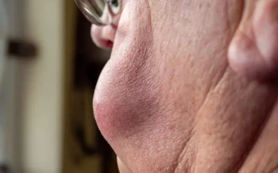
Regarded as one of the most serious types of skin cancer – Melanoma develops in the cells (melanocytes) that produce melanin – the pigment that gives your skin its color. Often resembling moles, melanoma growths occur when unrepaired DNA damage to skin cells (often caused by exposure to ultraviolet (UV) radiation from sunlight or tanning lamps and beds) triggers mutations (genetic defects) that lead the skin cells to multiply rapidly and form malignant tumors. These tumors originate in the pigment-producing melanocytes in the basal layer of the epidermis and can easily spread to other organs in the body. In most cases, melanomas appear as irregular brown, black and/or red spot. However, an existing mole can also begin to change color, size or shape. Avoiding sunburn is an effective way to reduce the risk of this type of skin cancer. Healthcare providers and medical coding companies must be thoroughly knowledgeable regarding the associated medical codes for melanoma.
According to reports from the Skin Cancer Foundation, an estimated 178,560 cases of melanoma (out of which – 87,290 cases will be in situ (noninvasive) and 91,270 cases will be invasive) will be diagnosed in the United States in 2018. Although melanoma accounts for only 3% of all types of the skin cancer, it has the highest death rate of all types and is more likely to spread (metastasize) in the body. The risk of melanoma is higher among women – in the age group 25 – 30 years. Identifying the warning signs of this condition at an early stage can ensure that cancerous changes are detected and treated before it spreads to other parts of the body, where it becomes hard to treat and can be fatal.
Common Signs and Symptoms
One of the most common signs of melanoma is the appearance of a new pigmented or unusual-looking growth on your skin or a change in the existing mole. These moles can appear in any part of the body that has exposure to the sun, such as your back, legs, arms and face. However, these can also occur in areas that don’t receive much sun exposure such as the buttocks, scalp, soles of your feet, palms of your hands and fingernail beds. These hidden melanomas are more common in people with darker skin. In most cases, melanomas have an irregular shape and are more than one color. Other signs and symptoms include –
- Skin changes, such as a new spot or mole or a change in color, shape, or size of a current spot or mole
- A skin sore that fails to heal
- A spot or sore that becomes painful, itchy, or tender, or which bleeds
- A spot or lump that looks shiny, waxy, smooth, or pale.
- A flat, red spot that is rough, dry, or scaly
However, melanoma doesn’t always begin as a mole. It can also occur on otherwise normal-appearing skin.
Types of Melanoma
Melanoma involves four different types –
- Superficial spreading melanoma – One of the most common types, this melanoma normally appears on the trunk or limbs. The cancerous cells tend to grow slowly at first, before spreading across the surface of the skin.
- Nodular melanoma – Generally, appearing on the trunk, head, or neck, this type grows more quickly and turns completely red as it grows.
- Lentigo maligna melanoma – Affecting older people, this type appears especially in those parts of the body that have been regularly exposed to the sun over several years.
- Acral lentiginous melanoma – A rare kind that occurs in people with darker skin, this condition appears on the palms of the hands, soles of the feet, or under the nails.
Factors that may increase your risk of melanoma include fair complexion, excessive ultraviolet (UV) light exposure, a family history of skin disease, having many or unusual moles and weakened immune system.
Making an Accurate Diagnosis and Effective Treatment
Making an early and accurate melanoma diagnosis is vital as this helps to find out whether the skin moles have spread to other areas and choose the best effective treatment option on that basis.
As part of the initial diagnosis, your physician will conduct a detailed head-to-toe inspection of your skin. A self-exam may also help patients to find out the moles, freckles and other skin marks that are normal and can notice specific changes (if any). If your physician suspects any specific spot that may be melanoma, a detailed biopsy will be done to confirm the same. As part of the biopsy, a pathologist will analyze the sample in detail wherein all or part of the suspicious mole or growth will be removed. Biopsy procedures used to diagnose melanoma include – Punch biopsy, shave biopsy and excisional and incisional biopsy. The type of biopsy procedure a patient needs to undergo will depend on several factors. Physicians will prefer to use punch biopsy or excisional biopsy to remove the entire growth (whenever possible). Incisional biopsy may be used when other techniques can’t easily be completed, such as if a suspicious mole is very large. After melanoma has been diagnosed, several other imaging tests like – Chest X-ray, Lymphoscintigraphy, Ultrasound, CT scan, MRI scan and PET scan may be recommended to find out if cancer cells have spread within the skin or to other parts of the body.
Once an accurate diagnosis of the skin condition is made, the next step is to determine the extent (stage) of the cancer. To determine the stage of cancer, physicians will check the thickness of melanoma (under a microscope and measure it with a special tool called – micrometer) and check whether it has spread to nearby lymph nodes (through a procedure known as a sentinel node biopsy). In addition, physicians will also check whether the skin over the area has formed into an open sore and whether any dividing cancer cells are found under a microscope.
The selection of treatment options for melanoma generally depend on several factors like – size and stage of cancer, overall health of the patient and their personal treatment preferences. For early-stage melanomas, surgical procedure will be considered the best option to remove the thin layer of tissue beneath the skin. On the other hand, if the cancer has spread beyond the skin, several other treatment options like – chemotherapy, radiation therapy, biological therapy, surgery to remove affected lymph nodes and medications (such as – Vemurafenib (Zelboraf), dabrafenib (Tafinlar) and trametinib (Mekinist) will be given to treat the advanced stage.
Dermatologists, oncologists, pathologists and other specialists providing treatment (that involves diagnosis, screening and other tests) for melanoma patients need to be adequately reimbursed for their services. The diagnosis must be carefully documented using the appropriate medical codes. Medical billing and coding services offered by experienced providers can help physicians ensure the correct codes for their medical billing purposes.
ICD – 10 Codes for Melanoma
C43 – Malignant melanoma of skin
- C43.0 – Malignant melanoma of lip
C43.1 – Malignant melanoma of eyelid, including canthus
- C43.10 – Malignant melanoma of unspecified eyelid, including canthus
C43.11 – Malignant melanoma of right eyelid, including canthus
- C43.111 – Malignant melanoma of right upper eyelid, including canthus
- C43.112 – Malignant melanoma of right lower eyelid, including canthus
C43.12 – Malignant melanoma of left eyelid, including canthus
- C43.121 – Malignant melanoma of left upper eyelid, including canthus
- C43.122 – Malignant melanoma of left lower eyelid, including canthus
C43.2 – Malignant melanoma of ear and external auricular canal
- C43.20 – Malignant melanoma of unspecified ear and external auricular canal
- C43.21 – Malignant melanoma of right ear and external auricular canal
- C43.22 – Malignant melanoma of left ear and external auricular canal
C43.3 – Malignant melanoma of other and unspecified parts of face
- C43.30 – Malignant melanoma of unspecified part of face
- C43.31 – Malignant melanoma of nose
- C43.39 – Malignant melanoma of other parts of face
C43.4 – Malignant melanoma of scalp and neck
C43.5 – Malignant melanoma of trunk
- C43.51 – Malignant melanoma of anal skin
- C43.52 – Malignant melanoma of skin of breast
- C43.59 – Malignant melanoma of other part of trunk
C43.6 – Malignant melanoma of upper limb, including shoulder
- C43.60 – Malignant melanoma of unspecified upper limb, including shoulder
- C43.61 – Malignant melanoma of right upper limb, including shoulder
- C43.62 – Malignant melanoma of left upper limb, including shoulder
C43.7 – Malignant melanoma of lower limb, including hip
- C43.70 – Malignant melanoma of unspecified lower limb, including hip
- C43.71 – Malignant melanoma of right lower limb, including hip
- C43.72 – Malignant melanoma of left lower limb, including hip
C43.8 – Malignant melanoma of overlapping sites of skin
C43.9 – Malignant melanoma of skin, unspecified
Avoiding excessive exposure to ultraviolet radiation is one of the major steps to reduce the risk of skin cancer. This can be achieved by avoiding sunburn during the middle of the day, wearing protective clothing, using sunscreen with a minimum sun protection factor (SPF) and avoiding tanning lamps and beds. In addition, patients need to be familiar with their skin tone and notice visible changes. Make it a habit to examine your skin regularly for new skin growth or changes in existing moles, freckles, bumps and birthmarks. People who work outdoors should take precautions to minimize exposure.
Medical billing and coding for skin conditions can be complex. By outsourcing these tasks to a reliable and established medical billing and coding company (that provides the services of AAPC-certified coding specialists), healthcare practices can ensure correct and timely medical billing and claims submission.


